
By Santa J. Bartholomew M.D. FAAP, FCCM
See Corresponding Journal Article: Multisystem Inflammatory Syndrome related to Covid (MIS-C)
A 9-year-old Asian boy was brought to emergency department on December 20th, 2021, with a typical presentation of MIS-C. He had an 8-day history of fever and a 5-day history of abdominal pain in the setting of having family members recently tested positive by PCR for COVID-19 on November 23rd, 2021.
His first symptom (day 0) was an intermittent “high grade” fever (the mother did not measure his temperature) associated with chills and daily intermittent mild sore throat, relieved with paracetamol.
On days 2–4, he developed mild intermittent periumbilical cramping pain and yellow watery stools without melena/hematochezia, which progressed to vomiting with anorexia. He had a 1–2 cm swollen area on his left posterior neck that spread to the right side over 2–3 days, it was not erythematous or tender.
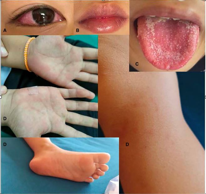
He had previously been brought to PCP and was diagnosed with tonsillitis and given amoxicillin without improvement. On day 5, he was more fatigued and developed bilateral nonexudative conjunctival injections, red lips, and increased anorexia. See pictures.
On day 6, the fever became persistent despite antipyretics. His abdominal pain, vomiting, and diarrhea worsened. His family brought him to an emergency department (ED). He was diagnosed with acute gastroenteritis without dehydration and was discharged with oral rehydration solution and analgesia/antipyretics and sent home
On day 7, he returned to PCP with the same symptoms not improving but with no change in diagnosis or treatment.
On the day of admission, day 8, his symptoms had not improved, and his family brought him to urgent care where he was diagnosed with suspected severe sepsis. At that time he had lymphopenia with an elevated white count , slightly raised CRP, normal abdominal ultrasound.
Subsequently, he was referred to the ED. His fever, fatigue, anorexia, and decreased urination continued, but the diarrhea decreased to once a day. He developed an occasional dry cough. He did not have any flank pain, urinary symptoms, other upper respiratory tract infection symptoms, chest pain, shortness of breath, change in senses of smell/anosmia, altered level of consciousness, headache, dizziness, visual disturbances, focal neurological symptoms, joint/muscle/bone pain or swelling, rash/eschar.
His mother and youngest sister had positive PCR results for COVID-19 three weeks prior. During that time, he had a low-grade fever and mild respiratory symptoms (runny nose and cough), but he was not tested for COVID-19. Two weeks before his illness, he and his family traveled to Vietnam, a vacation area. The family denied other recent travel.
Development: Normal
IUTD , no COVID vaccine
Medications: none
PSH: none
PMH: none
FH: non-contributory
At admission, his vital signs were temperature = 39.4 °C; respiratory rate = 50 bpm, heart rate = 150 bpm (regular), SpO2 = 95% on room air. His blood pressure was not recorded. His weight was 28.3 kg , and height was 124 cm. The patient was fatigued but alert, orientated, and cooperative. He had warm pink skin, capillary refill of < 2 s, and normal skin turgor. He had non-exudative conjunctival injection, red full lips, a white coating on his tongue, and itchy pink blanching maculopapular rashes at his palms, soles, and left knee. Non-tender, smooth, mobile enlarged lymph nodes were present in the bilateral posterior cervical chain (1.5–2 cm) and bilateral posterior triangles (left: 2 × 3 cm, right: 1.5–2 cm). There were no eschars, bruises, petechiae, desquamations, calf/muscle/joint/bone pain nor edema.
Front Pediatr. 2023; 11: 981880
He had mild intercostal retractions. He had regular S1 and S2 but with reduced heart sounds. He had no murmur nor gallop. His lung sounds were normal. He had mild abdominal distension with generalized tenderness on palpation, especially in the right upper quadrant, with mild to moderate guarding. There was no rebound, percussion, no flank tenderness. Bowel sounds were normal with no hepatosplenomegaly. The genital examination was not remarkable. He had no meningismus, the neurological examination was grossly normal.
On day 9 of illness, he clinically worsened and he was started on: intravenous immunoglobulin (IVIG at 2 g/kg in a single infusion over 12 h), methylprednisolone (2 mg/kg/day for 3 consecutive days), aspirin (3.5 mg/kg/day) ceftriaxone (100 mg/kg/day) for four total doses and Ceftrixaone and Clindamycin for presumed toxic shock syndrome.
Rest, oral nutrition, and oral hydration were also encouraged.
Twenty-four to 48 h after initiating IVIG and methylprednisolone, his clinical symptoms and fever improved. His heart and respiratory rates normalized, and oxygen therapy was no longer required. The lymphopenia and mild impaired cardiac function resolved. He was discharged from the hospital after 5 days with aspirin.
References
Case report: Managing multisystem inflammatory syndrome in children (MIS-C)
Vannida Douangboupha1,et.al.Front. Pediatr., 15 February 2023